|
Suture
This
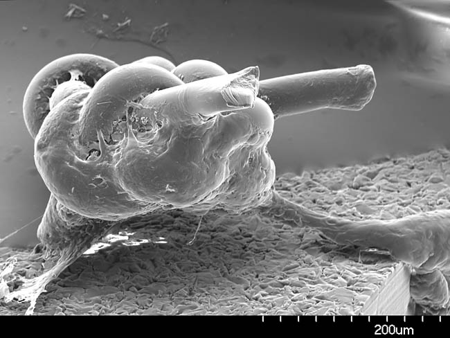
SEM micrograph shows a surgical suture.
A suture is basically a "stitch" created by a surgeon to repair tissue,
or to hold implanted devices in place. The suture material is
about 30 microns in diameter, and the overall knot is about 200 microns
in diameter. Creating such a small knot
requires a high degree of skill and dexterity on the part of a
surgeon.
It can be seen that a biofilm is beginning to
form in the crevices of the knot. At this magnification, it is not
possible to determine the constituents of the biofilm on the structure.
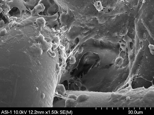
The image below shows a closer
magnification on the biomaterial encasing the suture.
This more detailed micrograph yields insight into some of the
constituents of the biomaterial forming on the suture.
Close up
View of Suture
The large cylindrical structure on the
left is the actual suture thread. There are indications of red
blood cells, which look like somewhat deformed donuts. The red blood
cells are slightly deformed, resulting from the sample preparation used
in creating the micrograph. In addition to red blood cells, there are
fibrous structures which are likely fibrin strands beginning a clotting
process. The more spherical structures in the lower center region of the
photograph look like they could possibly be some type of white blood
cells. The upper center portion of the micrograph shows an
indication of a bacterial biofilm forming. At this magnification, the
individual bacterium are barely visible, but close examination of the
photograph reveals a presence of a close packed array of spherical
cocci type bacteria. In other regions of the photograph it is hard to
distinguish between fibrin clotting agents and the adhesive matrix
excreted by the bacteria. |